Rabies virus was used for monosynaptic viral tracing. AAVs were used to label the morphology of specific neurons. (From
BrainVTA)
The viruses used in this article from BrainVTA are in the table below
|
Control |
PT-0473 AAV-CAG-Dio-mCherry |
|
Tracing Helper |
PT-0095 AAV-DIO-TVA-BFP
PT-0023 AAV-DIO-RG |
|
RV |
R01001 RV-△G-EnVA-EGFP
R01002 RV-△G-EnVA-DsRed |
Miao Ren, Jiaojiao Tian, Peilin Zhao, Jialiang Luo, Zhao Feng, Hui Gong, Xiangning Li
Pub Date: 2018-10-30,
DOI: 10.3389/fnins.2018.00885,
Email: [email protected]
Resin embedding has been widely used for precise imaging of fluorescently labeled biological samples with optical and electron microscopy. The low preservation rate of fluorescence, especially for red fluorescent proteins, has limited the application of resin embedding in multifluorescent protein-labeled samples. Here, we optimized the embedding method to retain the intensity of multiple fluorescent proteins during resin embedding. By reducing the polymerization temperature from 50 to 35 °C and adding a fluorescent protein protection reagent during the embedding process, we successfully increased the fluorescence preservation rate by nearly twofold for red fluorescent proteins, including tdTomato, mCherry and DsRed. Meanwhile, the background fluorescence decreased significantly in the optimized embedding method. This method is suitable not only for red fluorescent protein-labeled samples but also for blue (BFP) and green fluorescent protein (GFP)-labeled samples. We embedded brains labeled with BFP, DsRed and GFP via AAV and rabies virus and acquired the distribution of input neurons to different cortical areas. With GFP/tdTomato double-labeled samples in resin, we obtained the cholinergic projectome of the pedunculopontine tegmental nucleus (PPTg) and the distribution of cholinergic neurons at single-neuron resolution in the whole brain simultaneously. Input cholinergic terminals from the PPTg were found to innervate the cholinergic soma and fiber in the neocortex, basal forebrain and brainstem, indicating that local cholinergic neurons received long-range cholinergic modulation from the midbrain. Our optimized method is useful for embedding multicolor fluorescent protein-labeled samples to acquire multidimensional structural information on neural circuits at single-neuron resolution in the whole brain.
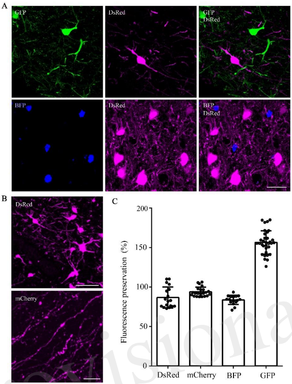
Figure 1. DsRed/mCherry/GFP/BFP can be well preserved using the optimized GMA embedding method.
In this study, the authors reported an optimized GMA-embedding method for multicolor fluorescent protein-labeled samples, which can retain over 95% of the fluorescence intensity of GFP, RFP (including tdTomato, DsRed, and mCherry) and BFP. This method can be used to acquire multi-structure information from BFP/GFP/RFP-labeled samples.
BrainVTA offers viral vector construction & virus packaging services for AAV, LV, RABV, PRV, HSV and VSV that help researchers explore questions about genes, neurons, circuitry structure, function of brain network, mechanism and treatment of diseases.
If you have any needs, just email us at
[email protected].