VSV
was used to couple with ferritin to generate a new trans-synaptic tracing tool.
The viruses used in this article are in the table below
|
VSV |
rVSV-dG-EGFP |
|
rVSV-dG-Ferritin-EGFP |
|
rVSV-EGFP |
|
rVSV-Ferritin-EGFP |
Pub Date: 2019-08-15,
DOI: 10.1016/j.neuroimage.2019.04.039,
Email: [email protected]
Ning Zheng, Peng Su, Yue Liu, Huadong Wang, Binbin Nie, Xiaohui Fang, Yue Xu, Kunzhang Lin, Pei Lv, Xiaobin He, Yi Guo, Baoci Shan, Anne Manyande, Jie Wang, Fuqiang Xu
The elucidation of neural networks is essential to understanding the mechanisms of brain functions and brain disorders. Neurotropic virus-based trans-synaptic tracing tools have become an effective method for dissecting the structure and analyzing the function of neural-circuitry. However, these tracing systems rely on fluorescent signals, making it hard to visualize the panorama of the labeled networks in mammalian brain in vivo. One MRI method, Diffusion Tensor Imaging (DTI), is capable of imaging the networks of the whole brain in live animals but without information of anatomical connections through synapses. In this report, a chimeric gene coding for ferritin and enhanced green fluorescent protein (EGFP) was integrated into Vesicular stomatitis virus (VSV), a neurotropic virus that is able to spread anterogradely in synaptically connected networks. After the animal was injected with the recombinant VSV (rVSV), rVSV-Ferritin-EGFP, into the somatosensory cortex (SC) for four days, the labeled neural-network was visualized in the postmortem whole brain with a T2-weighted MRI sequence. The modified virus transmitted from SC to synaptically connected downstream regions. The results demonstrate that rVSV-Ferritin-EGFP could be used as a bimodal imaging vector for detecting synaptically connected neural- network with both ex vivo MRI and fluorescent imaging. The strategy in the current study has the potential to longitudinally monitor the global structure of a given neural-network in living animals.
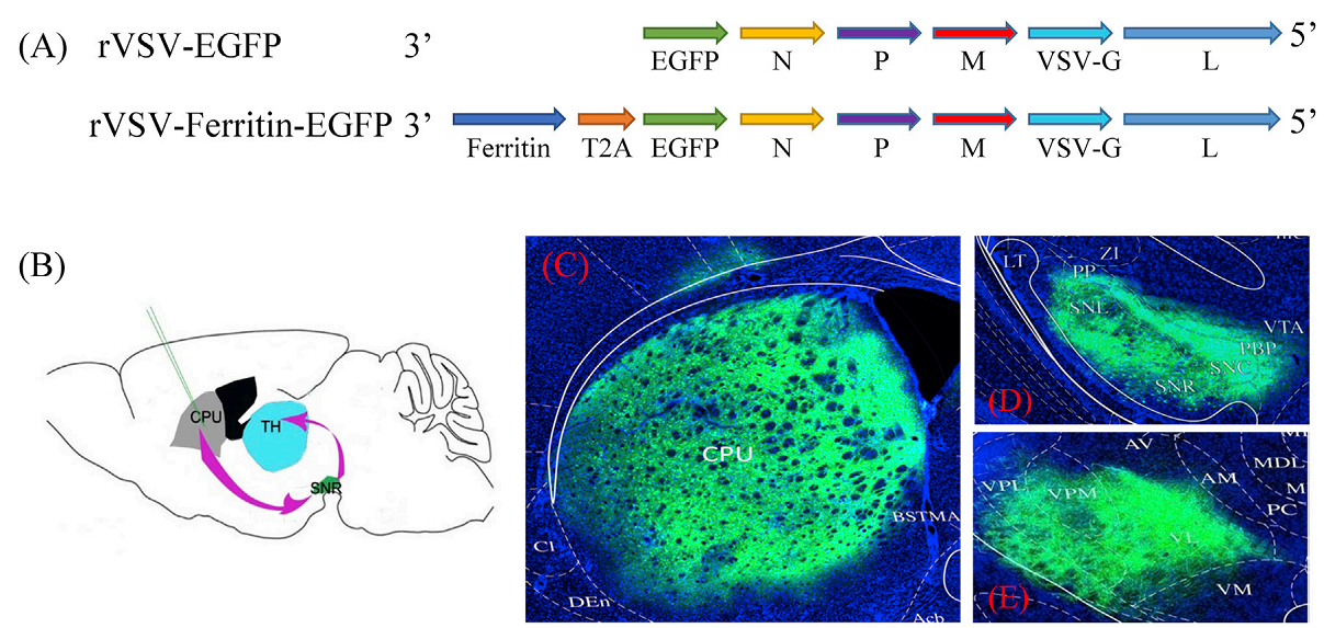
Fig1. rVSV-Ferritin-EGFP anterogradely transmitted across multiple synapses.
In this study, ferritin was coupled to enhanced green fluorescent protein (EGFP) gene and cloned into the VSV genome to generate a new trans-synaptic tracing tool. The results demonstrate that the structural neural connection can be detected with both MRI and fluorescent imaging. Thus, this study provides a novel strategy which combines the methods of neurotropic virus tracing and MRI to visualize the labeled global network for a given type or function in the brain, whose precise anatomical connections through synapses can be verified through optical microimaging.
BrainVTA offers viral vector construction & virus packaging services for AAV, LV, RABV, PRV, HSV and VSV that help researchers explore questions about genes, neurons, circuitry structure, function of brain network, mechanism and treatment of diseases.
If you have any needs, just email us at
[email protected].