VSV-EGFP and PRV-mRFP viruses (From
BrainVTA) were used for anterograde and retrograde tracing to evaluate whether disrupted neuronal circuits were repaired following NN tissue transplantation.
The viruses used in this article from BrainVTA are in the table below
|
Vesicular stomatitis virus |
VSV-EGFP |
|
pseudorabies virus |
PRV-mRFP |
Pub Date: 2019-09-19,
DOI: 10.1002/advs.201901240 Email: [email protected]
Bi‐Qin Lai, Ming‐Tian Che, Bo Feng, Yu‐Rong Bai, Ge Li, Yuan‐Huan Ma, Lai‐Jian Wang, Meng‐Yao Huang, Ya‐Qiong Wang, Bin Jiang, Ying Ding, Xiang Zeng, Yuan‐Shan Zeng
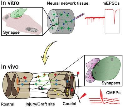
Tissue engineering produces constructs with defined functions for the targeted treatment of damaged tissue. A complete spinal cord injury (SCI) model is generated in canines to test whether in vitro constructed neural network (NN) tissues can relay the excitatory signal across the lesion gap to the caudal spinal cord. Established protocols are used to construct neural stem cell (NSC)‐derived NN tissue characterized by a predominantly neuronal population with robust trans‐synaptic activities and myelination. The NN tissue is implanted into the gap immediately following complete transection SCI of canines at the T10 spinal cord segment. The data show significant motor recovery of paralyzed pelvic limbs, as evaluated by Olby scoring and cortical motor evoked potential (CMEP) detection. The NN tissue survives in the lesion area with neuronal phenotype maintenance, improves descending and ascending nerve fiber regeneration, and synaptic integration with host neural circuits that allow it to serve as a neuronal relay to transmit excitatory electrical signal across the injured area to the caudal spinal cord. These results suggest that tissue‐engineered NN grafts can relay the excitatory signal in the completely transected canine spinal cord, providing a promising strategy for SCI treatment in large animals, including humans.
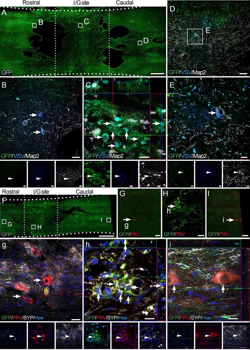
Fig.1 VSV anterograde and PRV retrograde tracing of transplanted NSC-derived neurons in the I/G site.
To evaluate whether disrupted neuronal circuits were repaired following NN tissue transplantation, VSV-EGFP and PRV-mRFP viruses(From
BrainVTA) were used for anterograde and retrograde tracing. The results showed that the preconstructed neural network tissue in vitro could establish synaptic connections with the nerve fibers transmitting the information of the descending brain nerve, and transmit the brain-derived excitatory nerve information to the motor neurons at the lower end of injury.
BrainVTA offers viral vector construction & virus packaging services for AAV, LV, RABV, PRV, HSV and VSV that help researchers explore questions about genes, neurons, circuitry structure, function of brain network, mechanism and treatment of diseases.
If you have any needs, just email us at
[email protected].